DSAEK Corneal Transplant Surgery in Huddersfield
Reliable Endothelial Keratoplasty for Fuchs' Dystrophy and Corneal Endothelial Failure
DSAEK (Descemet Stripping Automated Endothelial Keratoplasty) is a well-established partial-thickness corneal transplant that replaces the diseased endothelial layers with a precisely prepared posterior donor disc. It offers predictable outcomes, minimal external scarring, and is the preferred option in cases where greater technical reliability is required.
Affiliations & As Seen In


Read About Our Happy Patients
What a great experience! Very reassuring and I am very grateful for the consultation from Dr Musa he was so helpful in helping me make a decision to proceed to having eye correction surgery. I have the upmost confidence in him and the team at the Eye Doctor Clinic, Huddersfield.
Lozza 747I had implants with Dr Musa ten years ago. It was the best thing I've ever done. They are brilliant. My sight both near and distant was very poor but since the op I've had no glasses no lenses and my sight has been super. It remains really good to this day. Thank you Dr Musa. Brilliant consultations and treatment for glaucoma since.
Kay FitbitI can highly recommend the Eye Doctor Clinic, and I am so pleased I went there. Dr Musa, Gemma and Jess are lovely. They are very knowledgeable and were able to answer all my questions. My vision following surgery is great, and not having to wear glasses is fantastic.
Carol PeelWhat Is DSAEK?
DSAEK is a partial-thickness corneal transplant in which the diseased posterior stroma, Descemet membrane, and endothelium are replaced with a uniformly prepared donor disc, approximately 100–150 microns in thickness. The endothelium is the innermost cell layer responsible for keeping the cornea transparent and free of fluid; when it degenerates, the cornea swells and vision deteriorates.
Unlike full-thickness corneal transplantation, DSAEK preserves the patient's own anterior stroma and epithelium, leaving the majority of the cornea intact. The donor disc is delivered through a small incision, unfolded within the eye, and held against the recipient cornea by a sterile air bubble, which achieves natural adhesion within 24 to 48 hours.
DSAEK is a well-established alternative to DMEK in cases where graft handling presents greater technical complexity, including eyes with significant iris defects, vitrectomised eyes, or patients who are unable to maintain post-operative flat positioning. At The Eye Doctor, all DSAEK procedures are performed by Dr. Fayyaz Musa using quality-assured donor tissue and precision surgical technique.
Conditions Treated
- Fuchs' Endothelial Dystrophy: Genetic endothelial cell loss causing progressive corneal clouding and visual decline
- Bullous Keratopathy: Corneal swelling and blistering following cataract surgery or other intraocular procedures
- Failed Corneal Transplant: Re-grafting where a previous endothelial or full-thickness transplant has failed
- ICE Syndrome: Iridocorneal endothelial syndrome causing progressive unilateral endothelial loss
- Complex Cases: Eyes where DMEK presents higher technical risk, requiring a more manageable graft approach
When Is DSAEK Recommended?
Medical management may slow progression, but DSAEK is indicated when any of the following are present:
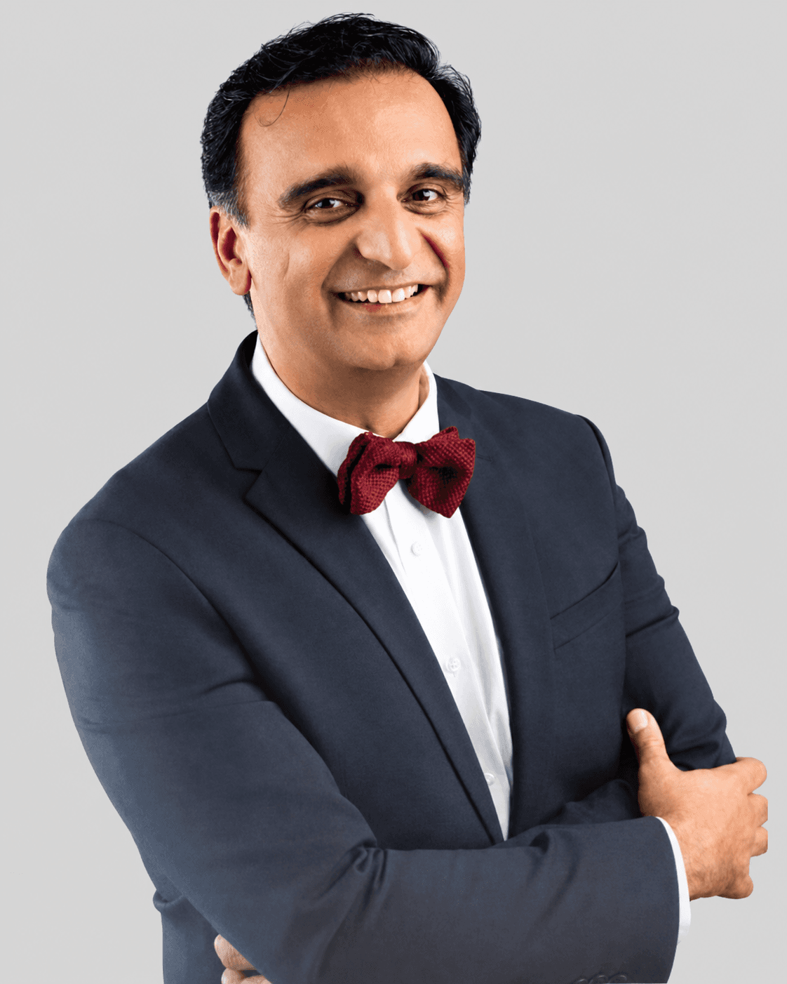
Professor Jeeva will assess corneal topography, endothelial cell count, and anterior segment imaging to determine whether DSAEK or an alternative technique is most appropriate for your presentation.
How DSAEK Surgery Works
The procedure is performed in two precise stages: automated graft preparation followed by delivery and air-bubble fixation.
Phase 1: Graft Preparation
Automated Donor Disc Preparation
- Step 1: Automated Cutting: A microkeratome creates a uniform posterior donor disc of approximately 100–150 microns thickness
- Step 2: Quality Assessment: The prepared graft is examined under specular microscopy to confirm endothelial cell density and tissue integrity
- Step 3: Recipient Preparation: The diseased endothelium and Descemet membrane are removed from the patient's cornea through a small incision
Phase 2: Implantation & Fixation
Graft Delivery, Positioning & Air Fixation
- Step 1: Graft Delivery: The donor disc is folded and carefully introduced into the anterior chamber through the small incision
- Step 2: Unfolding & Centration: The graft is unfolded and orientated precisely against the recipient posterior cornea
- Step 3: Air Fixation: A sterile air bubble is injected to press the graft into position; natural adhesion occurs over 24–48 hours
DSAEK at a Glance
Procedure Time
60–90 minutes
Anaesthetic
Local or general anaesthetic
Setting
Operating theatre, day case
Back to Work
2–4 weeks
Full Recovery
3–6 months
Surgery Cost
Contact us to inquire
Benefits of DSAEK Surgery
What to Expect During Surgery
Here is exactly what your experience will look like on the day of your procedure.
Most patients return home within a few hours of the procedure with a protective shield and written aftercare instructions.
Is This Treatment Right for You?
You may be a suitable candidate for DSAEK if you have:
A detailed pre-operative assessment will determine whether DSAEK or an alternative technique is the most appropriate choice for your condition and anatomy.
DSAEK may not be suitable if you have:
- Significant anterior corneal stromal scarring. Full-thickness keratoplasty may be more appropriate
- Active ocular infection or poorly controlled intraocular pressure. Must be treated before surgery
When to Consider Early Intervention
Earlier referral generally leads to better outcomes. A specialist assessment is recommended if you are experiencing any of the following:
Intervening before the cornea decompensates fully improves graft outcomes and shortens the visual recovery period.
Recovery After DSAEK Surgery
All post-operative drops, instructions, and follow-up appointments are arranged before you leave the clinic on the day of surgery.
DSAEK Surgery FAQs
Both DSAEK and DMEK are endothelial keratoplasty procedures, but they differ in graft thickness. DMEK transplants only the Descemet membrane and endothelium (10–15 microns), whilst DSAEK includes a thin layer of donor posterior stroma (approximately 100–150 microns). DSAEK is technically more forgiving to handle, making it the preferred choice in complex cases. DMEK typically produces marginally superior final visual acuity, but both techniques represent a substantial advance over full-thickness corneal transplantation.
Initial improvement is often noticeable within the first four to eight weeks. Full visual clarity typically develops over three to six months as corneal oedema resolves and the donor–recipient interface stabilises. Some patients with underlying conditions affecting other parts of the visual pathway may experience a more modest improvement, which Professor Jeeva will discuss openly at your consultation.
Rejection is a possibility with any form of corneal transplantation, though the risk with DSAEK is lower than with full-thickness penetrating keratoplasty. Any episode of sudden redness, pain, or visual decline should be reported promptly, as early treatment with intensive steroid drops can often reverse a rejection episode. Long-term low-dose steroid drops are prescribed to minimise this ongoing risk.
Partial graft detachment occurs in approximately 5–15% of DSAEK cases and is usually managed with a re-bubbling procedure in the operating theatre, in which air is re-injected to re-appose the graft. This is a straightforward intervention, typically performed under local anaesthesia, and does not usually compromise the long-term outcome.
Yes. Topical steroid eye drops are prescribed long term following DSAEK to reduce the risk of immune rejection. The frequency is gradually tapered over the first year and then maintained at a low maintenance dose. Regular follow-up appointments ensure that any pressure elevation from steroid use is identified and managed promptly.
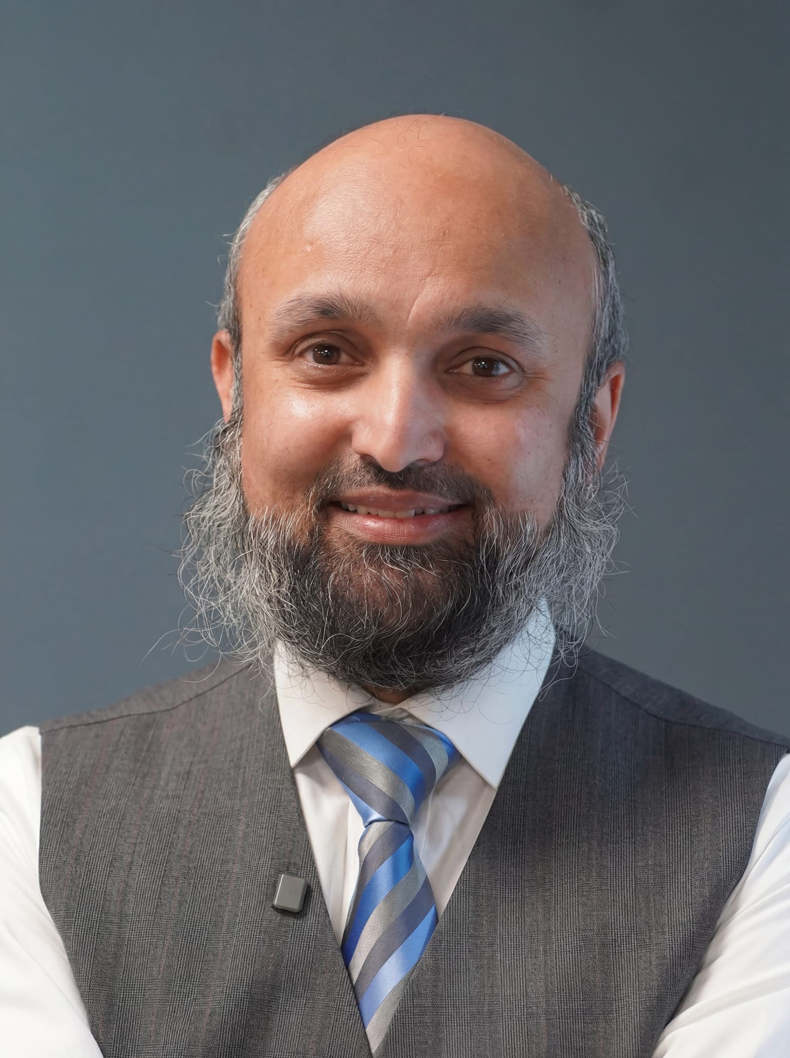
Dr Fayyaz Musa
MBChB (Edin) · FRCOphth (Lon) · CertLRS (RCOphth) · PGDipCRS
Founder & Consultant Corneal, Cataract & Glaucoma Surgeon

Dr Fayyaz Musa is the founder of The Eye Doctor Clinic and a recognised expert in anterior segment surgery. He is one of a small number of surgeons in the UK who holds dual fellowship training in both corneal disorders and glaucoma, and is the pioneer of DMEK surgery in the North of England. In cataract surgery, Dr Musa combines meticulous surgical technique with advanced biometry to deliver outstanding visual outcomes — including premium trifocal and toric lens implants for patients seeking full spectacle independence.
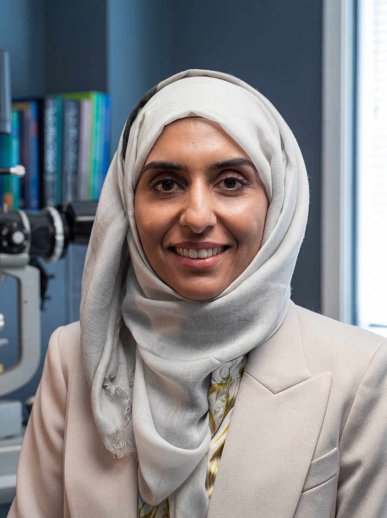
Meet the Team


Where to Find Us
Three convenient locations across West Yorkshire. Visit us for consultations, diagnostics, and treatments.
Bolton
136 – 140 Newport St
Bolton, Greater Manchester
BL3 6AB
Huddersfield
Woodlands, 4 Longbow Close
Huddersfield, HD2 1GQ
Consult a Specialist for DSAEK Corneal Transplant Surgery
A thorough assessment with Prof. Irfan Jeeva will confirm whether DSAEK is the right approach for you and what visual outcome you can realistically expect. Call +44 1484 627779 or book online.